Mole Screening Via Videodermatoscope By Dr. Rose Private Hospital In Budapest
- 3 May 2021 6:26 AM

 Malignant melanoma is an aggressive, cancerous skin tumor. With the help of annual screening tests, malignancies detected and removed in time are eminently curable, whereas in cases detected too late the malignant process can lead to metastasis.
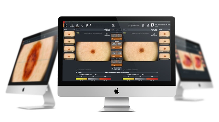
There are several melanoma screening methods available: beyond the classical dermatoscope, videodermatoscopic mole screening is now at the cutting edge. With the help of a videodermatoscope we can examine the whole body, any new skin formations, and any changes to existing moles.
By documenting changes in moles over time through comparison, we can filter out any skin formations that change as accurately as possible so that we can practically prevent a problem from occurring.
At Dr. Rose Private Hospital, we use state-of-the-art technology to track moles. During the examination, the whole body is photographed to thoroughly record each individual situation initially in a macro image, i.e., how many, and what size moles the patient has during the examination.
Each mole can be further examined with a so-called dermatoscopic probe, which allows us to magnify 10-20 or even 50 times.
With the help of magnification, we can see structures that help the doctor professionally, whether the skin formation is benign, questionable, or possibly malignant. This way, proper treatment can be started in time.
The analysis is also aided by other programs using artificial intelligence, drawing our attention to differences in size, shape, and structure, and thus confirming the doctor’s opinion on what to do next.
We have countless examples, in the event of a mole showing signs of growth that are nicely confirmed by high-resolution photographic documentation, whereby closer follow-up is required, in which case we recall the patient earlier than one year.
In this way it can be proved that the mole is benign, in which case no further action is required other than tracking any further changes. If there is a change in the structure, texture, and symmetry of the mole in addition to growth, surgical removal is recommended to prevent the development of melanoma.
Melanoma can develop without precedent, for example in the middle of a ’mole-free’ back, or it can develop in the process of changes in a pre-existing mole. For dermatological screenings, it is very important to have a mole examination once a year for everyone who is at an increased risk: for those of us with very light skin, red hair and a tendency to sunburn, the test is recommended twice a year.
This allows us to recognize a malignant change in a timely manner and provide a surgical solution as soon as possible before any malignant degeneration or even metastasis may develop.
For further information and to make an appointment, phone: +36 1 377 6737.
Click here to visit Dr. Rose Private Hospital online


LATEST NEWS IN business